Numbness or tingling in your shoulders back arms hands legs or feet. Your doctor may recommend an X-ray to look at the vertebrae surrounding a herniated disc.

Pin On Learning It S All Good
Regular X-rays will not show a herniated disc but they will give your doctor an idea of how much wear and tear is present in the spine and may show other causes of your problem.

. Your doctor may suggest taking X-rays of your lower back. While a standard X-ray cant show if you have a herniated disk it can show your doctor the outline of your spine and rule out whether your pain is caused by something else such as a fracture or. Often if a disc slips out of place the space between vertebrae may shrink or the vertebrae may become unstable without the disc to act as a cushion.
X ray only show pelvic tilt and L5S1 disc space narrowing. Compare Symptoms and Different Types of Back Pain and Sign Up for More Info. Ad Restore Your Back Health Get Lasting Pain Relief with Clinically Proven Technology.
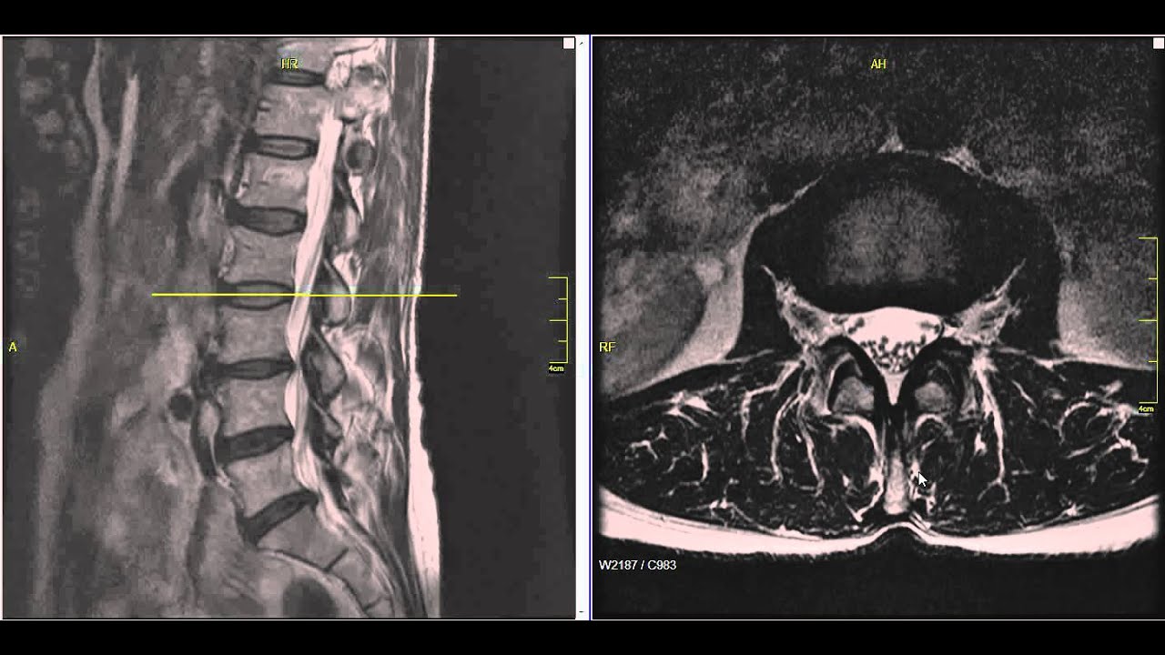
A slipped disc also called a prolapsed or herniated disc can cause. MRI actually show L34 disc herniation red arrow no L5S1 disc herniation yellow arrow that was suspected in X ray. X-rays use high-energy beams of light to create detailed images of the spine.
The most common test done today to diagnose a herniated disc is the MRI scan. Magnetic resonance imaging or mri scan report of spinal cord or lumbar bulging or lumbar vertebra magnetic resonance imaging or mri scan report of spinal cord or lumbar bulging x-ray Lumbar Spine Low Back lower disc bone problem bulging disc herniated disc slipped disc scan herniated disc x ray stock pictures royalty-free photos images. Ad Not All Back Pain is the Same.
Discs that become herniated usually are in an early stage of degeneration. Pain in the buttocks hips or legs if the disc is pressing on the sciatic nerve. Not Just The Symptoms.
Not all slipped discs cause symptoms. Located between each of the vertebra in the spinal column discs act as shock absorbers for the spinal bones. An X-ray a CT scan an MRI rarely a myelogram.
NeuroMD Is The Only Device That Corrects The Source Of The Pain. Because X-ray imaging can only show issues with the bones themselves and not the soft tissue a CT or MRI scan may be necessary to confirm the condition. Getting X-rays helps rule out other causes of back or neck pain.
MRI scan can diagnose whether the intervertebral disc is herniated and understand the condition of the spinal nerves. Problems bending or straightening your back. If they are dulled its often a sign of a herniated disc.
The doctor may tap just below the kneecap with a rubber hammer to see how the reflexes are working. This X-ray image includes the three lower lumbar vertebrae termed L3 L4 L5 and the sacrum S1 at bottom. Herniated disks can move into the space around your spinal cord and nerves and press on them.
Many people with lumbar disc herniations have dulled reflexes related to the patellar tendon. The doctor will confirm a diagnosis of lumbar disc herniation using X-ray imaging computed tomography CT scans andor magnetic resonance imaging MRI. Magnetic resonance imaging MRI scan of a sagittal section through the lumbar spine of a 52-year-old patient with a slipped herniated spinal disc white centre between the L2-L3 vertebrae associated with.
Degeneration of any of the intervertebral discs in the backbone is termed spondylosis. The intervertebral space between L4L5 is greatly reduced and indicates that the intervening disc has degenerated and has been partly squeezed out of the space between the. MRI can be used to diagnose disc disease.
Check if its a slipped disc. The most common and accurate imaging test for a suspected herniated disk is an MRI. Back and Knee Treatment Based on a Shoe.
See If An Underlying Source is Causing Your Pain. A CT scan show the bones of your spine. Slipped disc in spinal stenosis.
If your doctor thinks another condition is causing the pain or needs to see which nerves are being affected by the slipped disk they may order one or more of the following. Ad AposHealth Helps People with Knee Back and Hip Pain Live Well by Improving their Gait. Learn More About the Benefits of Apos.
Magnetic resonance imaging MRI. A herniated disc also called bulged slipped or ruptured is a fragment of the disc nucleus that is pushed out of the annulus into the spinal canal through a tear or rupture in the annulus. They may also check your reflexes muscle strength walking ability see if you can feel light touch pinprick vibration.
Lumbar Disc Herniation Mri Explained Dr Jeffrey P Johnson Hd Youtube Lumbar Disc Disk Herniation Degenerative Disc

Pin On Conditions

Pin Page

Neck And Back Degenerative Spondylolisthesis Conditions Spondylolisthesis Spine Surgery Lumbar Spinal Stenosis

The 5 Worst Exercises If You Ve Herniated Your Disc Livestrong Com

Unilateral Bilateral Locked Facets Cervical Spine Trauma Radiology Slip Spondylolesthesis Subluxation

Lateral Ct Scan Shows A Herniated Disc Protruding Into The Spinal Canal Cauda Equina Syndrome Cauda Equina Diagnostic Imaging

Disc Herniation Best Ways To Help Your Lumbar Disc Herniation

What S Another Option Instead Of Facet Joint Injections

Disc Replacement Vs Fusion

Pin On Bulging Disc

Pin On Back
What Core Exercises Can I Do With Degenerative Disc Disease And Herniated Discs Livestrong Com

Cauda Equina Syndrome From Lumbar Disc Herniation

Pivd

X Ray Image Two Views General X Ray And Mri Lumbar Spine Showing Herniated Nucleus Pulposus Of L4 L5 Intervertebral Disc Medical Image Concept Stock Photo

Pin On Mri

Pin On Chiropractic Works

Dr Gillard Lectures On How To Read Your Lumbar Mri
- soalan peperiksaan akhir tahun darjah 4
- peugeot 3008 ultimate red
- malaysia ron 95 price
- ocbc malaysia internet banking
- kata mutiara tabir kepalsuan
- gambar tali pinggang
- undefined
- slipped disc lumbar x ray
- usa olympic logo eps
- kartun budak kampung
- contoh resume terbaik 2016
- lagu hetty koes endang
- lukisan bendera malaysia berkibar
- kwsp i akaun majikan login
- jenis roti di roti o
- volkswagen cars in malaysia
- kalori ayam goreng spicy mcd
- tombiruo penunggu rimba full movie
- deko bilik tidur ikea
- gambar meja catok sablon